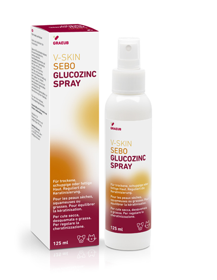
SCHUPPIGE UND FETTIGE HAUT BEI HUNDEN UND KATZEN

SCHUPPIGE UND FETTIGE HAUT BEI HUNDEN UND KATZEN

V-SKIN

SEBO

GLUCOZINC SPRAY

Zur Behandlung seborrhoischer Erkrankungen bei Hunden und Katzen muss die Haut mit Feuchtigkeit versorgt und einem sanften Peeling unterzogen werden. Die Hautbarriere muss wiederhergestellt und die Malassezia-Infektion bekämpft werden.

Das GlucoZinc-Spray dient der Wiederherstellung und Reparatur der Hautbarriere zur Aufrechterhaltung der Homöostase und des Ökosystems der Haut. Seine Anwendung ist zur Vorbeugung von Rückfällen hilfreich.

Es ist auch eine gute Ergänzung bei der Behandlung und Vorbeugung von Malassezia-Dermatitis.

Wichtige Punkte:

  • Enthält Gluconolacton, einen Bestandteil mit keratolytischer Wirkung für ein Peeling und die Reduzierung von Schuppenbildung.1, 2, 3, 4, 5
  • Mit der antibakteriellen und antimykotischen Wirkung von Zink.6
  • Mit Ceramiden zur Wiederherstellung des Epithels, zur Aufrechterhaltung der Barrierefunktion und zur Feuchtigkeitsversorgung der Haut.18, 19, 20

Anwendungsgebiet:

  • Leave-on-Spray, das auch in schwer zugänglichen Bereichen wie dem Interdigitalraum verwendet werden kann, um das Gleichgewicht der Haut wiederherzustellen und die Haut einem sanften Peeling zu unterziehen.

Art und Weise der Anwendung:

  1. Auf die gewünschten Bereiche aufsprühen.

Sehen Sie sich das Video zur Produktanwendung an!

Meinungen von Tierärzten

Was Sie über seborrhoische Haut wissen müssen

Literatur

  1. Berardesca E, Distante F, Vignoli GP, Oresajo C, Green B. Alpha hydroxyacids modulate stratum corneum barrier function. Br J Dermatol 1997;137:934-938.
  2. Hachem JP, Roelandt T, Schürer N, Pu X, Fluhr J, Giddelo C, Man M-Q, Crumrine D, Roseeuw D, Feingold KR, Mauro T, Elias PM. Acute acidification of stratum corneum membrane domains using polyhydroxyl acids improves lipid processing and inhibits degradation of corneodesmosomes. J Invest Dermatol. 2010;130:500-10.
  3. Bernstein EF, Brown DB, Schwartz MD, Kaidbey K, Ksenzenko SM. The polyhydroxy acid gluconolactone protects against ultraviolet radiation in an in vitro model of cutaneous photoaging. Dermatol Surg. 2004;30:189-196.
  4. Puigdemont A, Furiani N, De Lucia M, Carrasco I, Ordeix L, Fondevila D, Ramió-Lluch L, Brazis P. Topical polyhydroxy acid treatment for autosomal recessive congenital ichthyosis in the golden retriever: a prospective pilot study. Vet Dermatol. 2018; 29: 323-e113. May 22. doi: 10.1111/vde.12654
  5. Jarząbek-Perz, S., Mucha, P.; Rotsztejn, H. (2021). Corneometric evaluation of skin moisture after application of 10% and 30% gluconolactone. Skin Research and Technology, (5), 925–930.
    https://doi.org/10.1111/srt.13044
  6. Abendrot, M., & Kalinowska-Lis, U. (2018). Zinc-containing compounds for personal care applications. In International Journal of Cosmetic Science (Vol. 40, Issue 4, pp. 319–327). Blackwell Publishing Ltd.
    https://doi.org/10.1111/ics.12463
  7. Hekmatpou, D., Mehrabi, F., Rahzani, K., & Aminiyan, A. (2019). The effect of aloe vera clinical trials on prevention and healing of skin wound: A systematic review. In Iranian Journal of Medical Sciences (Vol. 44, Issue 1).
  8. Sánchez, M., González-Burgos, E., Iglesias, I., & Gómez-Serranillos, M. P. (2020). Pharmacological update properties of aloe vera and its major active constituents. In Molecules (Vol. 25, Issue 6).
    https://doi.org/10.3390/molecules25061324
  9. Teplicki, E., Ma, Q., Castillo, D. E., Zarei, M., Hustad, A. P., Chen, J., & Li, J. (2018). The effects of aloe vera on wound healing in cell proliferation, migration, and viability. Wounds, 30(9).
  10. Kamr, A., Arbaga, A., El-Bahrawy, A., Elsify, A., Khaled, H., & Hassan, H. (2020). The therapeutic efficacy of Aloe vera gel ointment on staphylococcal pyoderma in dogs. Veterinary World, 13(11).
    https://doi.org/10.14202/VETWORLD.2020.2371-2380
  11. Arbaga, A., El-Bahrawy, A., Elsify, A., Khaled, H., Hassan, H. Y., & Kamr, A. (2021). Biochemical and histopathological changes related to the topical application of Aloe vera ointment for canine pyoderma. Veterinary World, 14(5).
    https://doi.org/10.14202/vetworld.2021.1354-1362
  12. Preethia, K. C., Kuttanb, G; Kuttan, R. (2009). Anti-inflammatory activity of flower extract of Calendula officinalis Linn. and its possible mechanism of action. Indian Journal of Experimental Biology 2009 47(2)
  13. Tresch, M., Mevissen, M., Ayrle, H., Melzig, M., Roosje, P.; Walkenhorst, M. (2019). Medicinal plants as therapeutic options for topical treatment in canine dermatology? A systematic review. BMC Veterinary Researc, (1).
    https://doi.org/10.1186/s12917-019-1854-4
  14. Giostri, G. S., Novak, E. M.; Guarita-Souza, L. C. (2021). Treatment of acute wounds in hand with Calendula officinalis L.: A randomized trial. Tissue Barriers.
    https://doi.org/10.1080/21688370.2021.1994822
  15. Cruceriu, D., Balacescu, O; Rakosy, E. (2018). Calendula officinalis: Potential Roles in Cancer Treatment and Palliative Care. In Integrative Cancer Therapies (Vol. 16, Issue 4, pp. 1068–1078). SAGE Publications Inc.
    https://doi.org/10.1177/1534735418803766
  16. Lima, M. dos R., Lopes, A. P., Martins, C., Brito, G. A. C., Carneiro, V. C; Goes, P. (2017). The effect of Calendula officinalis on oxidative stress and bone loss in experimental periodontitis. Frontiers in Physiology, (JUN).
    https://doi.org/10.3389/fphys.2017.00440
  17. John, R.; Jan, N. (2017). Calendula Officinalis-An Important Medicinal Plant with Potential Biological Properties.Proceedings of the Indian National Science Academy,(0).
    https://doi.org/10.16943/ptinsa/2017/49126
  18. ung, J. Y., Nam, E. H., Park, S. H., Han, S. H.; Hwang, C. Y. (2013). Clinical use of a ceramide-based moisturizer for treating dogs with atopic dermatitis. Journal of Veterinary Science, 14(2), 199–205.
    https://doi.org/10.4142/jvs.2013.14.2.199
  19. Reiter, L. v., Torres, S. M. F.; Wertz, P. W. (2009). Characterization and quantification of ceramides in the nonlesional skin of canine patients with atopic dermatitis compared with controls. Veterinary Dermatology, 20(4), 260–266.
    https://doi.org/10.1111/j.1365-3164.2009.00759.x.
  20. Yoon, J. S., Nishifuji, K., Ishioroshi, S., Ide, K.; Iwasaki, T. (2013). Skin lipid profiling in normal and seborrhoeic shih tzu dogs. Veterinary Dermatology, 24(1).
    https://doi.org/10.1111/j.1365-3164.2012.01102.x.